



脳観

Description of 脳観
「脳観」は東京大学医学部脳神経外科にて開発された、脳幹の解剖とSafe entry zone (以下SEZ:脳幹へのアプローチとして安全とされる領域)を直観的に学習できるアプリです。
従来の教科書、解剖書は二次元的な表示に留まり、中脳、橋、延髄のそれぞれの断面を断片的に観察することでしか内部の神経線維、神経核を学習せざるを得ず、解剖のリアルな三次元的な空間把握はどうしても困難でしたが、自由自在に回転、拡大、透見可能な「脳観」で操作することでより主体的かつ直観的に三次元的な理解を可能にします。
脳幹の三次元モデルの開発に当たっては柿澤幸成医師(諏訪赤十字病院脳神経外科)が作成した3DCGをベースとしています。
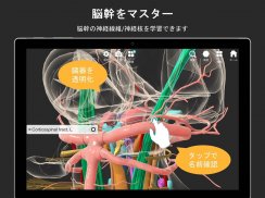
画面のドラッグやピンチで回転や拡大/縮小表示し、また解剖構造をタップして半透明/透明表示させ、内部の神経線維、神経核を表示します。
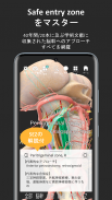
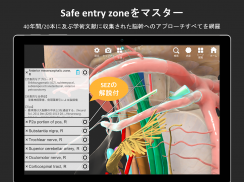
SEZはこれまでのPubMed上の文献20本(1982年~2019年)から情報を収集し、全部で14種類、24領域を表示しました。SEZをタップすることで重要な関連構造をハイライトし、代表的な手術アプローチ、合併症、そして文献から収集された関連情報を引用文献と共に表示します。
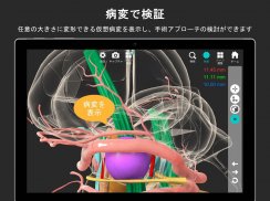
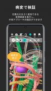
病変モードでは紫色の球体/楕円体で仮想病変を表示できます。この仮想病変は大きさ、位置を任意に設定でき、球体/楕円体の変形も可能です。例えば臨床現場にて実際の症例を模倣した病変を表示し、手術アプローチの検討を行うことが可能です。
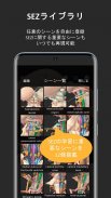
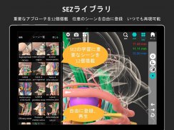
こうして検討されたシーンはシーン登録ボタンにてライブラリに保存でき、保存したシーンはシーン閲覧ボタンをタップすると瞬間的に呼び出すことができます。
"Brain view" is an app developed by the University of Tokyo Department of Neurosurgery that allows you to intuitively learn the anatomy of the brain stem and the safe entry zone (SEZ: a safe area for approaching the brain stem).
Conventional textbooks and anatomy books are limited to two-dimensional display, and internal nerve fibers and nerve nuclei can only be learned by observing the cross sections of the midbrain, pons, and medulla oblongata only in pieces. Although it was difficult to grasp the real three-dimensional space of, it is possible to freely and intuitively operate in a "brain view" that allows you to freely rotate, magnify, and see through a three-dimensional understanding. I will.
The development of the three-dimensional model of the brain stem is based on 3DCG created by Dr. Yukinari Kakizawa (Red Surgery, Suwa Red Cross Hospital).
Rotate or enlarge/reduce the display by dragging or pinching the screen, and tap the anatomical structure to make it semi-transparent/transparent and display the inner nerve fibers and nerve nuclei.
SEZ has collected information from 20 publications on PubMed (1982 to 2019) and displayed 14 types and 24 areas in total. Tap on the SEZ to highlight important relevant structures and view typical surgical approaches, complications, and relevant information gathered from the literature along with citations.
In lesion mode, virtual lesions can be displayed as purple spheres/ellipsoids. The size and position of this virtual lesion can be set arbitrarily, and the shape of a sphere/ellipsoid can be changed. For example, it is possible to examine the surgical approach by displaying lesions that mimic actual cases in the clinical setting.
The scenes examined in this way can be saved in the library with the scene registration button, and the saved scenes can be instantly called by tapping the scene browsing button.


